alan little’s weblog
my right knee, part two
2nd December 2004 permanent link
A story of hubris, yoga and the anatomy of the human knee. By request, and because Gwendoline Hunt makes an appearance in part three … Part Two: The Anatomy Of The Human Knee
In Part One: Hubris we left Alan in the summer of 1984, battered and broken on the ground at the bottom of a climb he really should not have been attempting that day. The broken left wrist healed quite quickly. The right knee, which had been diagnosed with strained ligaments, didn’t.
Fast forward ten years.
I could run. I studied karate for a few years with no knee trouble except that I had to be careful trying certain high side kicks. Climbing and hillwalking were no problem at all. But walking on pavements hurt. Anything over a mile or so and the knee started to swell and throb. Which itself, you might think, was not so terrible for a man – a good excuse not to go shopping. Better still, it was at its worst in cold, damp autumn weather. Praise the lord, no Christmas shopping.
But it wasn’t funny, in fact, and it wasn’t getting any better. More alarming: there was a lump growing out of the side of the knee that was so hard it felt like bone. I wanted to be able to walk around town, and I didn’t want extra bones growing out of the side of my knee. Unfortunately the wonderful British health service doesn’t give a damn about patients whose problems aren’t (yet) crippling. Several attempts to get doctors to take an interest in “doctor, my knee aches if I walk on pavements a lot” failed. At one point I had a GP [family doctor] who was a runner himself and should have known something about taking knee injuries seriously, but he didn’t give a damn either.
I decided, finally, that I was going to have to do something about it. A friend in my climbing club said he knew a good sports physiotherapist who worked with the national kayak team and would definitely take injuries from a climbing accident seriously, even if they were by this time ten years old. I booked an appointment. I arrived. “Take your trousers off and sit on the couch, please”, he said. He was doing something at his desk, several metres away on the other side of the room. He turned round.
“You’re going to need surgery for that”.
Closer inspection confirmed the several-metres-away instant diagnosis. The hard lump was a cyst that had probably formed around a tear in the outer edge of the meniscus. It was forcing the outer edge of the knee joint apart – but the serious problem, he said, was the inner edge of the joint which was therefore being pinched together. If it wasn’t dealt with I would have arthritis within five years. He arranged for me to see his friend, a top knee surgeon, who agreed and booked me in for an arthroscopy. An arthroscopy is one of those small-scale knee operations where they insert a fibre optic camera through a tiny incision, have a look what’s actually going on in there and fix it with tiny miniature instruments and minimal trauma.
I woke up. Sure enough, I had two tiny, barely visible incisions, one on each side of the knee. And a big three inch scar up the outside of my leg, where they had decided the tiny miniature instruments were futile and attacked the cyst with industrial scale digging and drainage equipment. At least it didn’t hurt.
What they don’t tell you after a knee operation is that the general anaesthetic may have worn off, but your leg is still stuffed full of local painkillers. After a few hours they wear off too. Then it hurts.
After a while it stops hurting. It seemed like a good idea to get back to moving and using the leg as quickly as possible, so I was swimming within a few weeks and climbing again – carefully – within a couple of months. And the knee was somewhat better, the swelling was gone. But autumn came, and it still ached.
I went back to the original physiotherapist. This time he referred me to his partner who was the team physio for Manchester City Football Club, and therefore knew a lot about knees (kayakers, I presume, are better at hurting their shoulders). His theory was this: there is a structure called the ilio-tibial band, which is a strip of tendon that runs down the outside of the leg and stops the knee from collapsing outwards. The surgeon would have had to cut through the ilio-tibial band in order to get to the cyst in my knee; the physio suspected the problem I now had was that the scar in the ilio-tibial band hadn’t healed well and was possibly compressing some nerves in that area. If that was the case, though, he said there honestly wasn’t much he could do for me. He gave me a course of ultrasound treatment, some general exercises to strengthen and stabilise my knee, and taught me some stretches that were supposed to work on the ilio-tibial band to some degree. They maybe helped a bit.
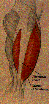
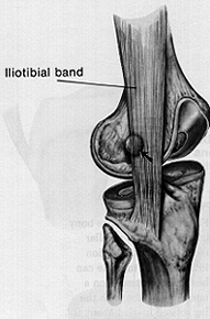
Pictures courtesy of a website about iliotibial band friction syndrome – which is apparently a common problem for runners, but it’s not what I had
And there we left it. I had had the best treatment that western surgery and sports physiotherapy had to offer. My knee was somewhat better than it was before, and livable with but by no means perfect. It was better than having arthritis at the age of forty, but still quite disappointing really.
related entries: Yoga
all text and images © 2003–2008